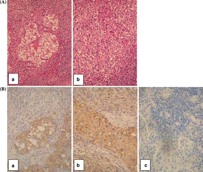
Fig. 6.
H&E and immunohistochemical staining of a RCC lesion from which the RCC52 cell line was established.a H&E staining shows the mixed clear cell (central area) and sarcomatoid RCC (a) and sarcomatoid RCC representing a majority of the tumor population (b).b Immunostaining with the β 2m-specific mAb L368 resulted in the staining of clear cell component, but did not stain or barely stained the sarcomatoid component of the tumor tissue (a). Immunostaining with the HLA class I heavy chain-specific mAb HC-10 resulted in moderate to strong staining of the surface and cytoplasm of the clear cell component and in weak to moderate staining of the cytoplasm of the sarcomatoid component of the tumor tissue (b). No staining of the tumor tissue section was detected with normal mouse IgG (NMIgG) (c). Original magnification, ×400

