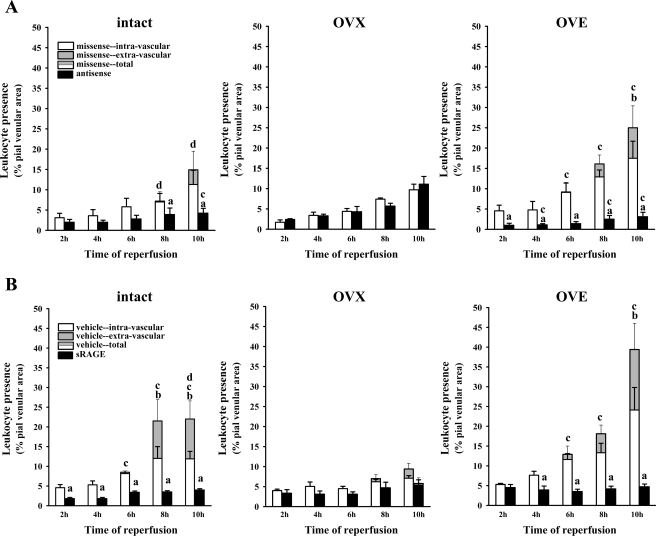
Fig. 5.
A: postischemic leukocyte behavior (over 2–10 h reperfusion), following 48 h exposure to topically applied RAGE antisense or missense ODN, in diabetic intact [n = 5 (antisense), and n = 4 (missense)], OVX [n = 6 (antisense), and n = 7 (missense)], and OVE [n = 7 (antisense), and n = 7 (missense)] females. The data are expressed as the percentage of the viewed venular area occupied by adherent (i.e., “nonrolling”) leukocytes present within the intravascular compartment [missense (white bars) or antisense (black bars)] or as the area percentage of leukocytes found extravascularly (gray-shaded portion of bars only, seen solely in missense-treated rats). Total leukocyte presence (intravascular + extravascular leukocytes) is represented by the combined height of the gray/white bars. The SE bars arising from the tops of the gray portions of the data bars relate to total leukocyte presence. The SE bars originating from the tops of the white data bar portions relate to intravascular leukocytes. Statistical comparisons (P < 0.05) at each time point: aintravascular adhesion-antisense vs. missense, btotal vs. intravascular leukocyte presence, cintact or OVE total missense or antisense vs. OVX, and dintact vs. OVE total leukocyte presence. B: postischemic leukocyte behavior at 24 h following intracerebroventricular injection of sRAGE or artificial cerebrospinal fluid vehicle in diabetic intact, OVX, and OVE females (n = 4 in all cases, except OVE + sRAGE, where n = 6). The data are expressed as the percentage of the viewed venular area occupied by adherent leukocytes present within the intravascular compartment [vehicle (white bars) or sRAGE (black bars)] or as the area percentage of leukocytes found extravascularly (gray-shaded portion of bars only, seen solely in vehicle-treated rats). Substituting “vehicle” for “missense” and “sRAGE” for “antisense,” SE bar descriptions and statistical symbol definitions are the same as in A.

