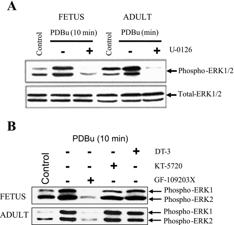
Fig. 3.
A: Western immunoblots of p-ERK1/2 levels at 10 min in fetal and adult cerebral arteries in response to PDBu (3 × 10⁻⁶ M) in the absence or presence of the MEK inhibitor U-0126 (10−5 M). B: Western immunoblot of PDBu-mediated increase (at 10 min) in p-ERK1 (p44) and p-ERK2 (p42) in fetal and adult cerebral arteries in the presence of the PKG inhibitor DT-3 (2.5 × 10⁻⁷ M), the PKA inhibitor KT-5720 (10−6 M), and the PKC inhibitor GF-109203X (5 × 10−6 M).

