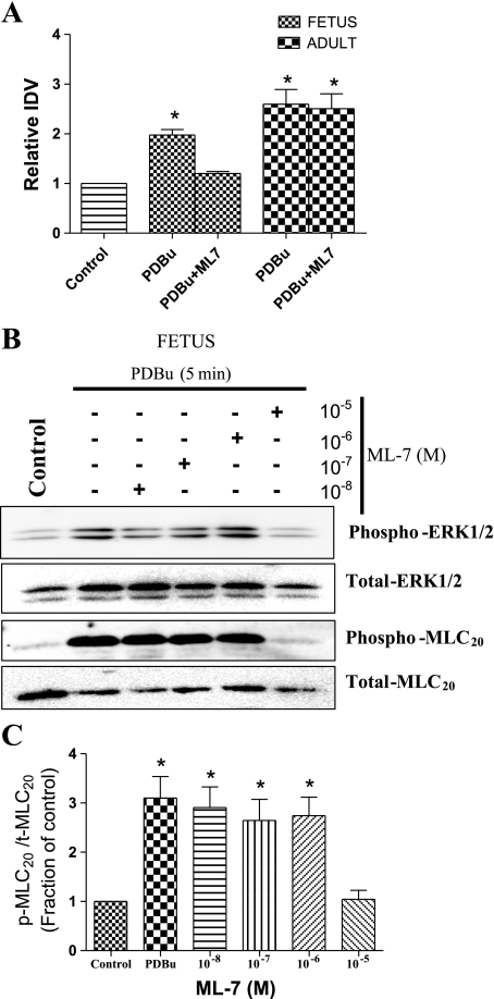
Fig. 6.
Role of myosin light chain kinase (MLCK) in PDBu-mediated MLC20 activation in the fetal and adult cerebral arteries. A: densitometric analysis of MLC20 phosphorylation in response to 3 × 10⁻⁶M PDBu alone and 3 × 10⁻⁶M PDBu + 10−5 M ML-7 in the fetal and adult cerebral arteries as detected by Western immunoblot analysis. IDV, integrated density values. B and C: Western immunoblots (B) and densitometry analysis as means ± SE (C) of ML-7 concentration-response relations for p-MLC20 in presence of PDBu in fetal cerebral arteries incubated with combinations of various concentration (10−8 M to 10−5 M) of MLCK antagonist, i.e., ML-7 for 30 min before stimulation with PDBu (3 × 10⁻⁶ M). *P < 0.05.

