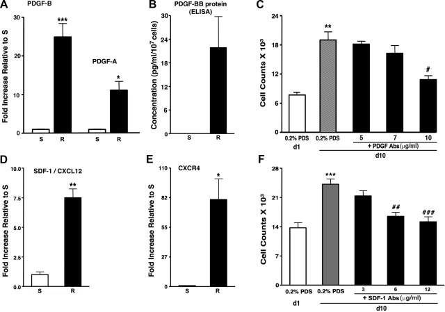
Fig. 5.
Autocrine growth of dR cells is due, in part, to PDGF-BB and SDF-1/CXCL12. A: real-time PCR analysis demonstrates that dR cells express higher levels of mRNA for PDGF-B and PDGF-A than dS-SMC (26.4 ± 1.6-fold and 13.2 ± 1.3-fold, respectively). Specific messages were normalized to HPRT. Data are presented as fold-change of relative gene expression in dR cells compared with that of dS-SMC. ***P < 0.0003, *P < 0.05. B: dR cells secrete PDGF-BB at concentrations of 21.84 ± 7.92 pg/ml/107 cells, as detected by ELISA in serum-free medium conditioned by cells for 72 h (samples from 5 dR cell populations were analyzed). S-SMC do not make PDGF-BB at detectable levels by ELISA (samples of conditioned medium from 3 dS-SMC populations were analyzed). C: autocrine, serum-independent growth of dR cells (gray bar) is partially attenuated by neutralizing PDGF-BB/AB antibodies (added at 5, 7, 10 ng/ml). Cell counts were performed on day 1 (d1) and day 10 (d10). **P < 0.05 compared with d1 cell counts; #P < 0.05 compared with d10 untreated (0.2% PDS) cell counts. D and E: real-time PCR analysis demonstrates that dR cells express higher levels of mRNA for SDF-1/CXCL12 (D) and its receptor CXCR4 (E) (10.0 ± 1.2-fold and 80.84 ± 19.19-fold, respectively) compared with dS-SMC. Specific messages were normalized to HPRT. Data are presented as fold-change of relative gene expression in dR cells compared with that of dS-SMC. **P < 0.005, *P < 0.05. F: autocrine, serum-independent growth of dR cells (gray bar) is attenuated by neutralizing SDF-1/CXCL12 antibodies (added at 3, 6, 12 ng/ml) in a dose-dependent manner. ***P < 0.0005 compared with 0.2% PDS; ##P < 0.005 and ###P < 0.001 compared with the effect of R-CM without SDF-1 antibodies.

