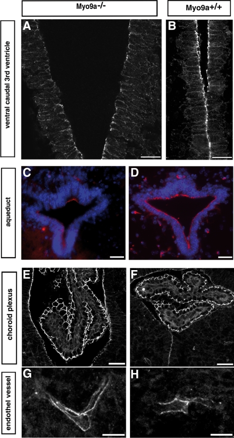
Figure 4.
Occludin staining is decreased in tight junctions of ependymal epithelium of the ventral caudal 3rd ventricle and the aqueduct, but not in the epithelium of the choroid plexus or the endothelium lining the blood vessels. Immunofluorescence staining of coronal sections from P0.5 Myo9a−/− (A, C, E, and G) and WT littermate mice (B, D, F, and H) for occludin showing that it is reduced in tight junctions of the ependyma in the ventral caudal 3rd ventricle (A) and the aqueduct (C, red) from Myo9a−/− mouse brains. No difference in occludin staining was observed in the epithelium of the choroid plexus (E and F) and the endothelium lining the blood vessels (G and H) in Myo9a−/− compared with control tissue. Scale bars, (A and B) 20 μm; (C–H) 30 μm.

