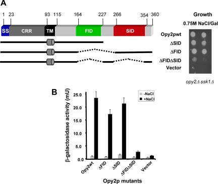
Figure 3.
Two functional regions of the Opy2p cytoplasmic tail. (A) Schematic representation of deletion constructs of the Opy2p cytoplasmic tail (left), assayed in yeast cells (opy2Δ ssk1Δ) by serial dilution spotting assay for their ability to grow on hyperosmolarity media (right). (B) The capacity of activating the HOG pathway by yeast cells (opy2Δ ssk1Δ) bearing different mutants assayed using a transcriptional reporter (8xCRE-CYC1-LacZ) assay.

