Figure 5.
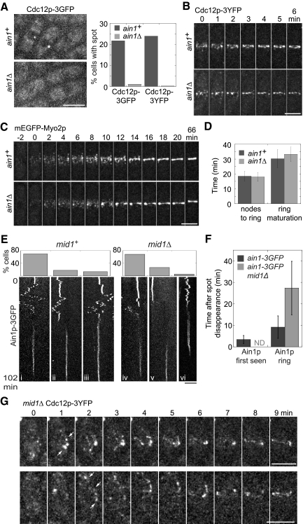
The contractile ring assembles from nodes in the absence of the spot (A–D) and the spot is dispensable for the node-independent ring assembly pathway (E–G). (A) Localization of Cdc12p to the spot depends on Ain1p. Left, micrographs of maximum intensity projections of 23 sections spaced at 0.2 μm. Right, percentage of ain1+ (strains JW1404 and JW1405) and ain1Δ (JW1456 and JW1722) interphase cells with Cdc12p spots (n > 250 cells each). (B) A time series of ring assembly from Cdc12p-3YFP nodes in ain1+ and ain1Δ cells. Maximum intensity projections of six slices spaced at 0.8 μm resulting in fewer detectable nodes and less homogeneous ring appearance. (C) Time series showing ring assembly from nodes in the absence of a spot (strains JW1109 and JW1136). Maximum intensity projections of nine sections spaced at 0.6 μm. Node appearance is set as time zero. (D) The timing of node condensation into a ring and ring maturation was similar in ain1+ and ain1Δ cells expressing mEGFP-Myo2p (JW1109 and JW1136). The same cells were analyzed at both stages: ain1+, n = 15; ain1Δ, n = 25. (E) Graphs (top) and kymographs (bottom) of Ain1p-3GFP showing the relationship between spot movement (top portion of the kymographs) and contractile-ring formation (bottom portion of the kymographs) in mid1+ and mid1Δ cells. Kymographs were constructed using a slit parallel to the long axis of maximum intensity projections of the cells. Z-sections were collected at 0.8-μm spacing every 30 s for 102 min (see Materials and Methods). The cells from mid1+ (n = 23, JW1149) and mid1Δ (n = 18, JW1450) were grouped into three categories (graphs show the percentage of cells in each): (i and iv) The Ain1p spot moved to the division site and disappeared before ring formation; (ii and v) The Ain1p spot disappeared distant to the division site before ring formation (see Video 9); (iii and vi) The spot was integrated into the contractile ring. (F) Temporal relationship (mean ± SD) between Ain1p-3GFP spot disappearance, the first sign of Ain1p-3GFP accumulation in the contractile ring and the appearance of a sharp Ain1p ring in mid1+ (n = 20) and mid1Δ cells (n = 17). ND, not determined. (G) The Cdc12p filament and ring do not originate from a spot in most mid1Δ cells. Strain JW1189 (mid1Δ cdc12-3YFP) was observed for 30 min. Cdc12p-3YFP appears at several locations near the division site (white arrows) in two representative cells at time 1 min. The signals at 0 min are cytoplasmic speckles. Bars, 5 μm.

