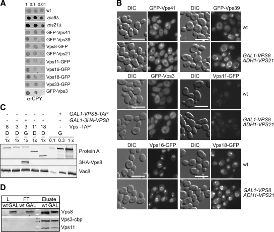
Figure 5.
HOPS and CORVET subunit distribution on the Vps21 compartment. (A) CPY sorting. The indicated strains were spotted on YPD plates and analyzed as in Figure 4F. (B) Localization of GFP-tagged Vps41 and Vps39 (top), Vps3 and Vps11 (middle), and Vps16 and Vps18 (bottom) upon Vps8 overproduction. Fluorescence microscopy analysis was performed as in Figure 1A. (C) Relative expression levels of tagged CORVET subunits. Cells expressing the indicated TAP-tagged proteins were grown in glucose and galactose. Protein extracts were analyzed by SDS-PAGE followed by Western blotting. Extracts from GAL1-VPS8-TAP cells were loaded in the indicated dilutions to the gel. (D) CORVET assembly. C-terminally TAP-tagged Vps3 was purified form strains carrying HA-tagged Vps8 under the control of either the VPS8 or the GAL1 promoter using IgG beads. Load (L), flow-through (FT), and eluate were analyzed by SDS-PAGE and Western blotting using antibodies directed against Vps11, the calmodulin-binding peptide (cbp) or the HA tag.

