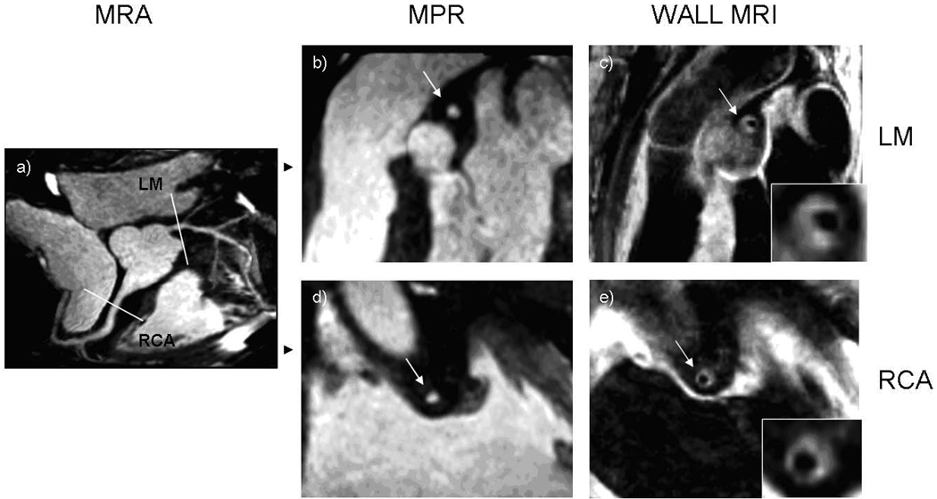
Figure 1. 73 year old male participant with increased wall thickness and preservation of lumen area.
(a) Magnetic resonance angiogram (MRA) shows no significant stenosis in the proximal portions of the coronary arteries. Multiplanar reconstruction (MPR) of the MRA of the left main (b) and the right (d) coronary arteries shows a normal lumen in cross-sections. Coronary wall MRI of the corresponding arterial cross-sections (c, e) shows eccentrically thickened arterial walls. Mean wall thickness: 4.1 mm for the left main, 2.5 mm for right coronary artery. Lumen and outer contour areas 7.1 mm2 and 54.3 mm2 for the left main and 7.0 mm2 and 51.4 mm2 for the right coronary artery.

