
Figure 1.
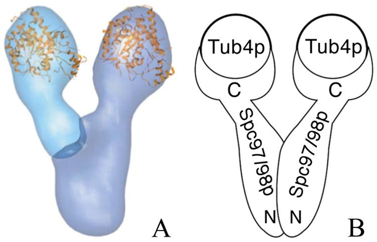
A) Electron microscopy reconstruction of the S. cereviseae γ-TuSC. Light blue represents the flexibly attached arm and the more rigid body is presented in dark blue. The location of γ-tubulin was indicated by gold labeling, and the crystal structure of human γ-tubulin was manually fit into the structure (Kollman et al. 2008). B) A schematic representation of the arrengement of γ-TuSC proteins based on the relative orientations of N- and C- termini determined by in vivo FRET.