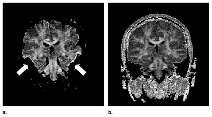
Figure 2.
Susceptibility-related signal loss and eddy-current artifacts commonly affect the single-shot diffusion-weighted EPI images. On the anisotropy maps, areas corresponding to regions of low signal on the diffusion-weighted image have been thresholded (white arrows) and eddy-current related image distortions give rise to borders with high anisotropy values (white arrows). Fractional anisotropy maps obtained with LSDI (right side) are relatively free of such artifacts.

