
Figure 4.
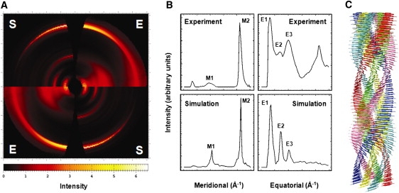
Experimental and simulated diffraction patterns of an insulin amyloid fibril and a six-protofilament β-roll-based insulin fibril model. (A) Quadrant view of the experimental insulin x-ray fiber diffraction pattern (E) (26) and the simulated fiber diffraction patterns of a six-protofilament insulin fibril model using the β-roll monomeric subunit as a building block, constructed by DISORDER (S) (48). The image was prepared by FIT2D (36). (B) Meridional and equatorial arc profiles of the experimental and simulated diffraction patterns, with two meridional arcs, at ∼9.6 Å and ∼4.8 Å (M1 and M2, respectively), and three equatorial arcs, at ∼33 Å, ∼15 Å, and ∼10 Å (E1–E3, respectively). (C) Tentative model of the six-protofilament β-roll-based insulin amyloid fibril. The image was prepared by the UCSF Chimera package (48). The experimental x-ray fiber diffraction pattern of insulin amyloid was collected under the experimental conditions of the SAXS analysis from which the mature fibril consisting of six intertwining protofilaments was evaluated (26).