
Fig. 3. Defect of calvarial ossification in Icap-1 mutant mice.
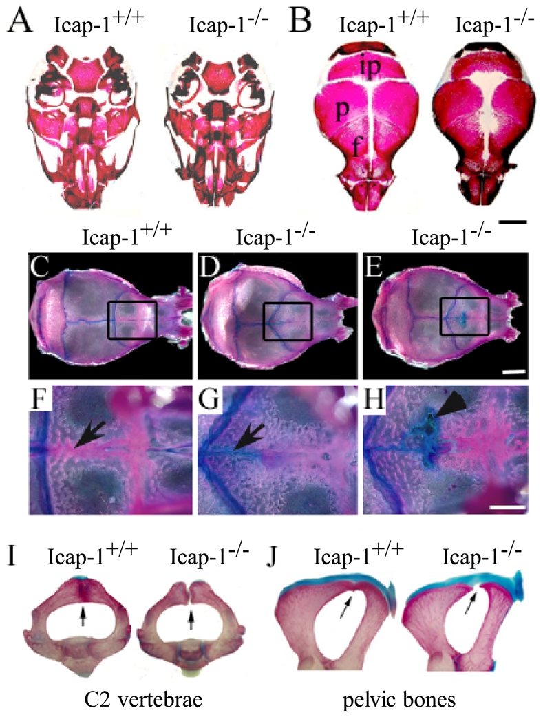
(A–B) Whole-mount alizarin red staining of the skulls of Icap-1+/+ and Icap-1−/− newborn mice. (A) Mineralization of the skull base is comparable in wild-type and mutant animals. (B) Mineralization of the skull vault. The mineralized areas of the interparietal (ip), parietal (p) and frontal bones (f) are smaller and fontanelles are open in Icap-1−/− mice. Bar, 2 mm. (C-H) Whole-mount alizarin red/alcian blue staining of wild-type and Icap1-null 2-month-old calvariae. The area of the metopic suture is boxed in C-E, and displayed at high magnification in F-H. In Icap1-null (D, E) the sagittal suture is shorter, the coronal sutures are V-shaped and irregular when compared with wild-type litter mates (C). At this age, the metopic suture (arrow) is closed in wild-type (F), whereas in Icap-1-deficient mice the posterior part of the metopic suture is not ossified and stained with alcian blue (G, H). Some Icap-1−/− mice display non-mineralized, alcian blue-positive areas in the frontal bones (arrowhead in H). Bars are 5 mm (C–E) and 2 mm (F–H), respectively. (I, J) Whole-mount skeletal staining of the axis (I) and the pelvic region (J) of 21-days-old wild-type (wt) and Icap-1−/− (mt) mice. Arrows point to the fusion defect observed in the Icap-1-deficient mice while fusion is completed in control animals.