
Fig. 5. Reduced proliferation of calvarial osteogenic cells in Icap-1 null mice.
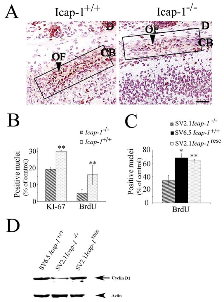
(A) Immunodetection of the proliferation marker Ki67 in the sagittal sutural region of newborn Icap-1+/+and Icap-1−/− calvaria. The osteogenic front of the Icap-1−/− mice displays a reduced number of Ki67-positive cells. Boxes indicate regions used for KI-67 and BrdU quantification in (B). Bar is 25 μm. (B) Quantification of Ki67- and BrdU-positive cells in the osteogenic front of control and mutant animals. Error bars represent S.D. Asterisks indicate a statistically significant difference between Icap-1+/+and Icap-1−/− (**, P<0.0001). (C) Immortalized calvarial osteoblasts. ICAP-1-deficient cell (SV2.1-Icap-1−/−) show significantly reduced BrdU-labeling index compared to wild-type cells (SV6.5-Icap-1+/+). Retroviral transfection of the Icap-1 cDNA into the Icap-1−/− cells rescues the proliferation defect (SV2.1-Icap-1resc) (**, P<0.0001). (D) SV2.1 and SV2.1-Icap-1resc cells were cultured for 24 h in 1% FCS before replating them onto 10 μg/ml FN. After 5 h of spreading, cells were washed with PBS and directly lysed onto Petri dishes with RIPA buffer. An amount of 30 μg of proteins per lane was gel separated and then transferred onto PVDF membrane before processing for Western blotting with the anti-cyclin D1 antibody. The same gel was blotted with anti-actin polyclonal antibodies for normalizing protein loading.