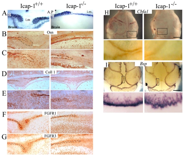
Fig. 6. Osteogenic differentiation is abnormal in Icap-1−/− calvaria.

Frontal sections through the parietal bones and the sagittal suture of Icap-1+/+ and Icap-1−/− newborn mice were stained for (A) AP, (B, C) osteonectin, (D, E) Col1 (F) FGFR1 and (G) FGFR3. (C, E) are (400X) magnification views of (B) and (D) osteogenic front region, respectively. Dashed lines represent bone borders and osteogenic front area. B, bone; i.m., intersutural mesenchyme. Note the reduced expression of the osteogenic and differentiation markers in Icap-1−/− tissue.
Whole mount in situ hybridization on calvaria of E17.5 embryo was performed to detect either Cbfa1/Runx2 (H) or Bsp transcript (I) on Icap-1+/+and Icap-1−/− embryos. The top panel is an overview of the full calvaria, the bottom panel is a closer view of the osteogenic front (boxed in the overview). Bars are 2 mm.
