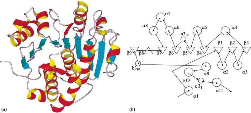
Fig. 2.
Ribbon drawing and topology of the subunit structure of YXKO. (a) The ribbon drawing of the YXKO subunit structure showing the overall fold. β strands are presented as blue arrows, α helices are depicted as red and yellow coils, and loops are shown in gray. (b) Topology diagram of the YXKO fold (α helices are shown as circles and β strands as triangles).

