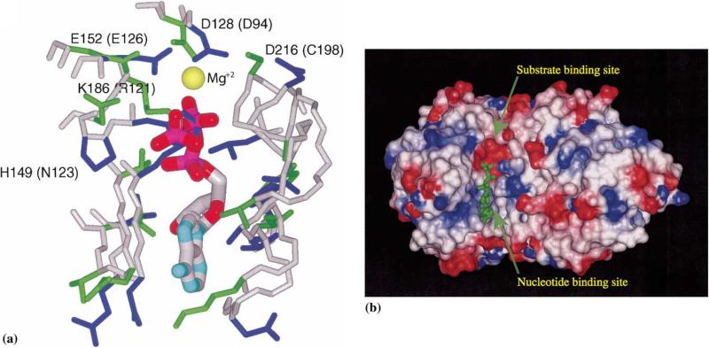
Fig. 4.
The putative ATP-binding site in YXKO. (a) The superposition of the YXKO structure onto ThiK, with ATP originating from ThiK; magnesium ion conserved within the family is indicated as a yellow sphere; the side chains of residues involved in the interactions with ATP are blue (YXKO) and green (ThiK); the names of ThiK residues corresponding to YXKO residues are in parentheses. (b) The putative ATP- and substrate-binding sites in YXKO, with van der Waals surfaces colored according to the molecular electrostatic potential on the residues: blue indicates positive potential, red indicates negative potential.

