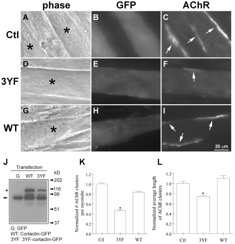
Figure 5. Inhibition of agrin-induced AChR clustering by forced expression of phospho-mutant cortactin in myotubes.
To examine the effect of exogenous cortactin proteins on AChR clustering, C2 myotubes were transfected with mRNAs encoding GFP (Ctl) or GFP-tagged phospho-mutant (3YF) cortactin or wild-type (WT) cortactin. After treating myotubes with agrin overnight, cells expressing exogenous proteins (A, D, G; asterisks) were identified by green fluorescence (B, E, H) and the AChR clusters present on the surface of these cells were examined by R-BTX-labeling (C, F, I; arrows). Forced expression of the phospho-mutant, but not wild-type, cortactin reduced the number and lengths of agrin-induced AChR clusters in myotubes. J. To biochemically confirm the expression of exogenous cortactin proteins in myotubes, extracts prepared from myotubes transfected with mRNAs encoding GFP, GFP-tagged WT and 3YF cortactin were immuno-blotted with anti-cortactin monoclonal antibody mAb4F11. Myotubes transfected with GFP mRNA (G) contained full-length endogenous cortactin (arrow on left), but those transfected with WT- and 3YF-cortactin mRNAs contained endogenous cortactin plus a protein (∼25 kD larger) corresponding to exogenous, GFP-tagged cortactin (asterisk). MW marker positions are indicated on the right. K-L. Myotubes transfected with GFP or GFP-tagged cortactin proteins were selected randomly and the numbers and lengths of the AChR clusters present on their surface were determined; data from five separate transfection experiments were pooled and normalized relative to values obtained from GFP-tranfected cells. Fewer (K) and smaller (L) AChR clusters were present in myotubes expressing phospho-mutant cortactin than in cells expressing GFP alone or WT-cortactin-GFP. Mean and SEM values are shown, *P<0.05.

