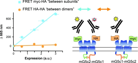
Figure 3. Metabotropic glutamate receptors form strict dimers at the cell surface.
mGlu5 subunits carrying the C-tail of GABAB1 (mG5c1 in orange, not reaching the cell surface alone) or GABAB2 (mG5c2, in green, allowing targeting to the cell surface of the C1-C2 combination), and taged with a myc and HA epitope, respectively, were labelled with FRET-compatible anti-HA antibodies only, or with a combination of anti-HA and anti-myc antibodies. Experiments were performed with cells expressing different amounts of receptors at the cells surface as quantified by ELISA. Note that a large TR-FRET emission, proportional to the amount of cell surface receptors, is observed when both subunits of the dimer are labelled. In contrast, no TR-FRET can be measured if only one subunit per dimer is labelled. Adapted from Kniazeff et al. (2004a).

