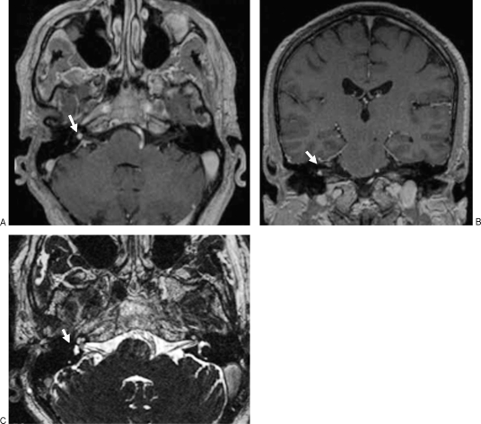
Figure 5.
Follow-up magnetic resonance (MR) images obtained 3 months after surgery. Axial (A) and coronal (B) T1-weighted MR images with contrast shows persistent contrast-enhancing lesion in the fundus of the internal auditory canal (arrows). (C) Axial 3D-constructive interference in steady state (CISS) image demonstrates that the tumor obstructs the cochlear fossa on the right side (arrow).

