Figure 3.
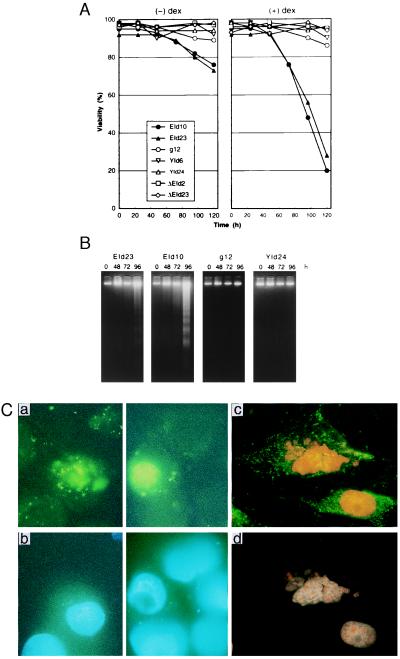
Induction of apoptosis in EId10 and EId23 cells after coexpression of E1A12S, Id-1H, and Id-2H. (A) Cell viability. The cells were cultivated in the presence and absence of 1 μM dex. Cell viability determined by trypan blue exclusion is expressed as the percentage of the total cell number. (B) DNA fragmentation. DNA was isolated from both floating and adherent cells by the method of Hirt and subjected to electrophoresis. DNA was stained with ethidium bromide. (C) Annexin analysis of apoptotic cells. Sparse cultures of EId10 cells on coverslips were treated with dex for 72 h and 100 ng⋅ml−1 of hoechst 33342, a permeable DNA-specific fluorochrome was added to the culture for 15 min. The cells then were stained with annexin-V fluorescein (Boehringer Mannheim) and PI, an impermeable fluorochrome, in the absence (a and b) and presence (c and d) of 0.1% Triton X-100, which renders the cells permeable. Photographs were taken under a fluorescence microscope at a magnification of ×1,000 with the excitation (λex) between 455 nm and 500 nm (a and c) and with the λex between 330 nm and 450 nm (b and d). Annexin-V fluorescein shows the localization of phosphatidylserine at the outer layer of the plasma membrane (a) and at both inner and outer layers (c). Hoechst-stained nonpermeabilized cell DNA (b) and both Hoechst- and PI-stained permeabilized cell DNA (d).

