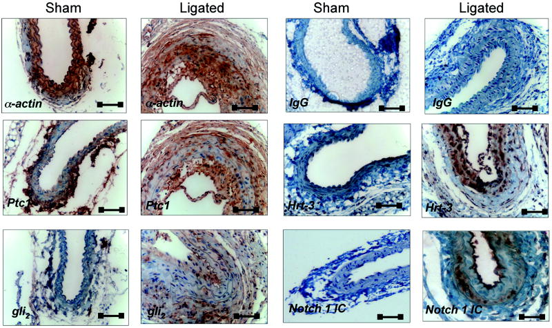
Figure 4. Hedgehog components in murine SMC in vivo.
(a) Hemotoxylin and eosin staining of a carotid artery (CA) from C57Bl6/J mice 14 d post ligation and (b) photomicrographs of immunohistochemical staining for SMC α-actin, Notch 1 IC, Ptc1, Hrt-3 and Gli2 in carotid arteries 14 d post ligation. Magnification 40x. Scale bars=50 μm.


