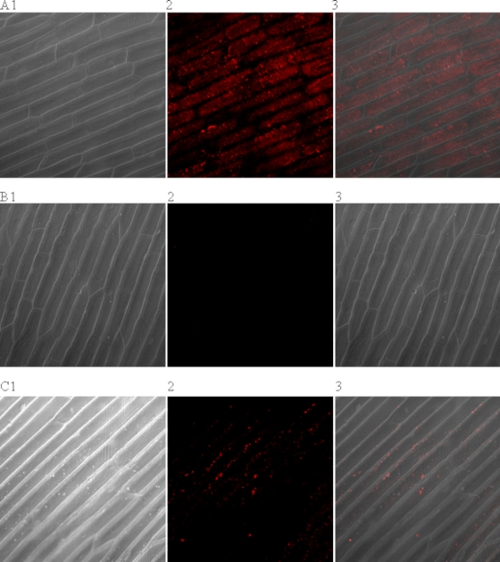
FIG. 2.
Visualization of biotinylated virus binding to the surface of onion epidermis. (A) Onion epidermis after exposure to bio-HAV and Q-Dots 655. (B) Onion treated with the Q-Dots only. (C) Onion epidermis after the bio-HAV had been eluted from the surface with beef extract buffer before the addition of the Q-Dots. Image 1 shows the onion epidermis only under light microscopy, image 2 shows the fluorescence from the Q-Dots, and image 3 is an overlay of images 1 and 2.

