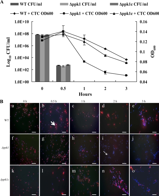
FIG. 2.
Effect of ppk1 deletion on the formation of VBNC cells of C. jejuni. (A) Culturability of the wild-type, Δppk1, and Δppk1c strains after formic acid treatment as determined by CFU enumeration and OD600 measurements after CTC staining. The data represent the mean ± SE of three independent experiments. (B) Fluorescence microscopy images of CTC-DAPI-stained C. jejuni cells showing viable (CTC-stained red cells, thick arrow) and dead (DAPI-stained blue cells, thin arrow) cells at different time points after formic acid treatment. Microscopy images a, b, c, d, and e (wild-type strain); f, g, h, i, and j (Δppk1 mutant strain); and k, l, m, n, and o (complemented strain) are the representative images of three independent experiments treated with formic acid for 0, 0.5, 1, 2, and 3 h, respectively. Note the drop in viable cells (red cells) of the Δppk1 mutant at 1, 2, and 3 h posttreatment compared to the wild-type strain. Bars, 10 μm. WT, wild type.

