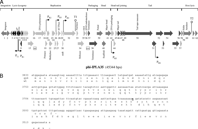
FIG. 3.
Physical and genetic map of the bacteriophage phiIPLA35. (A) The ORFs are sequentially numbered, indicated by arrows proportional to their lengths and pointing toward their direction of transcription (L = left, R = right). Some ORFs have been placed below for clarity. The functional modules are indicated on top of the scheme, and the names of several putatively or experimentally identified genes are shown below. Promoter (P) and terminator (T) sequences are also indicated. (B) DNA sequence of gp6 showing the insertion mutation (in bold and underlined) and alignment of the putative CI amino acid sequence of the mutant and the wild-type (wt) phages, respectively. Numbers to the left of the DNA sequence are the genome coordinates.

