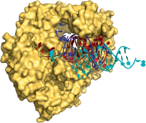
Figure 1.
Structure of Tgo-Pol bound to a uracil-containing primer-template (red) (13). Superimposed are the expected positions of DNA bound in the polymerization mode (cyan) (17) and the editing mode (blue) (18) derived from structures of the family-B polymerase from bacteriophage RB69. The position of the uracil-containing primer-template clearly maps to the editing conformation more closely than to the polymerization.

