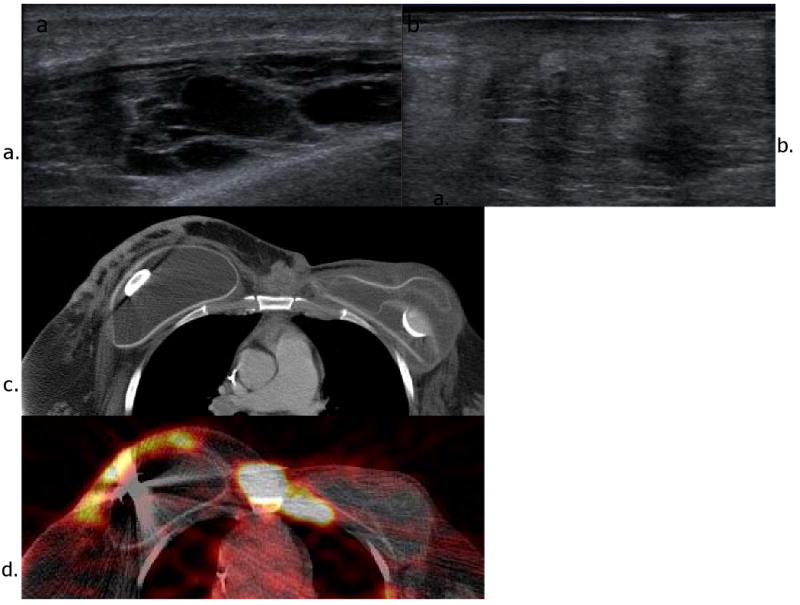
Figure 1.

Recurrence in the contralateral breast in a patient with history of left breast cancer and left partial mastectomy 10 years ago. She also underwent left total mastectomy and prophylactic right mastectomy when she had recurrence 6 years ago. Ultrasound images (a and b) show multiple hypoechoic and heterogeneous mixed echogenic lesions with shadowing in the right breast. Axial CT (c) and FDG PET-CT images (d) reveal hypermetabolic soft tissue mass and nodules with irregular skin thickening in the right breast.
