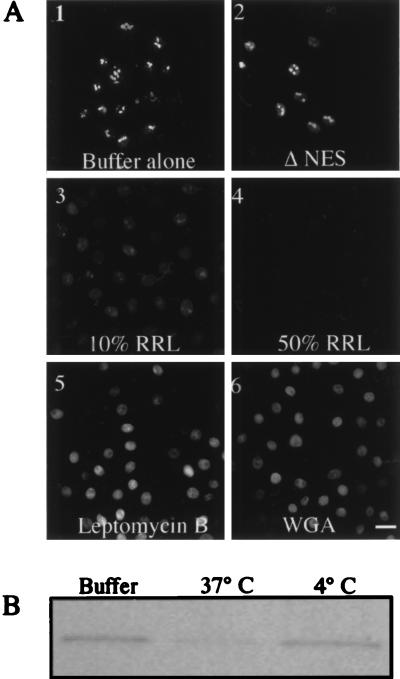
Figure 3.
Reconstitution of nuclear export in vitro. (A) Nuclear export was examined in vitro in RGG2.2 cells by fluorescence microscopy. After incubation with 1 μM corticosterone at 37°C, for 30 min, the cells were washed and permeabilized with 40 μg/ml of digitonin. Permeabilized cells then were incubated either in buffer A (1) or with transport buffer containing 50% RRL (2–6) with and without indicated inhibitors. A-2 represents cells transiently expressing a Rev/Gr/GFP chimera containing the M10 mutation. All cells were visualized by confocal laser microscopy. (Bar = 25 μm.) (B) Immunoblot analysis of nuclear export in vitro. Rev/Gr/GFP was detected in total cellular lysates from cells incubated in buffer A (Buffer) and cells incubated in transport buffer containing 50% RRL at indicated temperatures.

