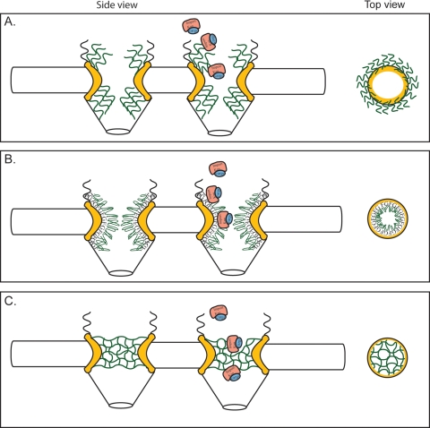
FIG. 3.
Models for the mechanism of NPC selectivity and transport. Based on the different features of the respective models, the distribution and physical features of the FG domains are distinct. This is represented in structural models of both a side view (perpendicular to the NE) and top view (cross-section through center of NPC; e.g., from cytoplasm onto plane of NE). NE, black; FG domains, green; structural NPC elements, yellow; importing karyopherin transport receptor, pink; NLS-bearing cargo, blue. (A) Brownian virtual gating model (138). The center of the NPC is a narrow channel, from which FG domains extend to form an entropic barrier to transport. Transport receptors bind these FG domains, overcoming the entropic barrier. By collecting on the NPC periphery, transport complexes increase the probability that they will spontaneously move across the barrier. (B) Reduction of Dimensionality model (125, 126). FG repeats form a continuous surface along the inner face of the NPC, and transport complexes pivot along this surface. The spacer sequence between FG repeats loop outward, forming a physical barrier to diffusion of large molecules; transport complexes might transiently displace these as they move along the FG surface. (C) Selective phase-partitioning model (133, 134). Hydrophobic interactions between FG repeats form a physical meshwork with gel-like properties. Transport receptors bind and transiently dissolve the meshwork in order to translocate through the NPC.

