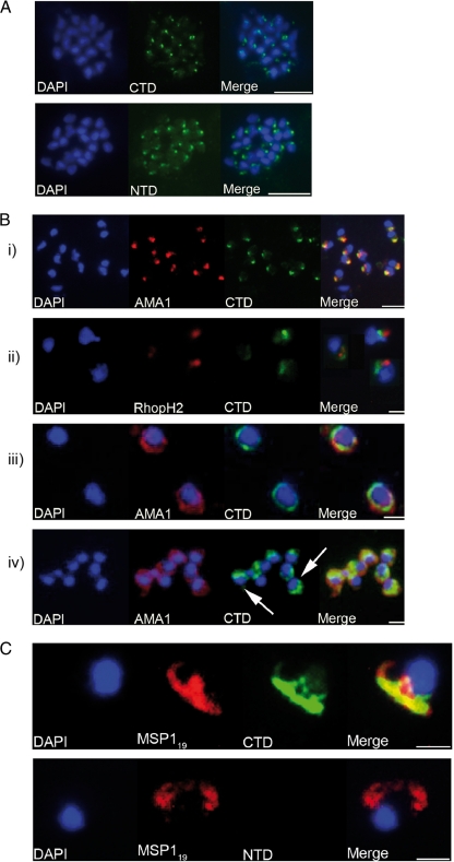
FIG. 5.
Analyses of the distribution of GAMA in asexual blood stage parasites by immunofluorescence. (A) Formaldehyde-fixed P. falciparum 3D7 schizonts were probed with mouse anti-CTD (top panel) or anti-NTD (bottom panel) antiserum and an anti-mouse-fluorescein isothiocyanate conjugate (green). Parasite nuclei were stained with DAPI (blue). Staining with both antisera resulted in a strong punctuate pattern, with a single point of fluorescence located away from the nucleus and consistent with a location in the apical organelles. Scale bars represent 2 μm. No signal was detected when parasites were probed with normal mouse serum (not shown). (B) Formaldehyde-fixed free P. falciparum merozoites were probed with rabbit anti-CTD antiserum and either mouse polyclonal anti-AMA1 antibodies (panels i, iii, and iv) or mouse MAb 61.3 (panel ii). Mouse antibodies were detected with an anti-mouse Alexa Fluor 594 conjugate (red) and rabbit antibodies with an anti-rabbit Alexa Fluor 488 conjugate (green). Parasite nuclei were stained with DAPI (blue). Scale bars are 5 μm in panel i and 2 μm in the other panels. In panels i and ii there is a clear apical localization of AMA1, RhopH2, and GAMA, with AMA1 and GAMA appearing to be colocalized; panel iii shows a circumferential localization of both GAMA and AMA1 on the surface of free merozoites; and panel iv shows a “cap”-like distribution of GAMA on the surface of free merozoites (arrows), in contrast to the circumferential distribution of AMA1. (C) Newly invaded ring stage parasites were probed with the mouse anti-MSP119 MAb 1E1 and either rabbit anti-CTD (top panel) or anti-NTD (bottom panel) antiserum. Mouse antibodies were detected with an anti-mouse Alexa Fluor 594 conjugate (red) and rabbit antibodies with an anti-rabbit Alexa Fluor 488 conjugate (green). Parasite nuclei were stained with DAPI (blue). There was no reactivity exhibited by mouse preimmune serum with ring stage parasites (not shown). Scale bars are 2 μm. There is a clear reactivity of anti-CTD antibodies, but not anti-NTD antibodies, with ring stage parasites. Anti-CTD and anti-MSP119 signals appear to colocalize (top panel), suggesting that the “stub” part of GAMA that is produced upon shedding of the p42-p37 complex is carried into the host cell and remains associated with the ring stage parasite.

