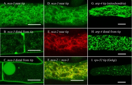
FIG. 5.
NCA-3 localizes to the tubular vacuolar network and the plasma membrane. The mitochondrial marker, ARG-4, and the Golgi marker, VPS-52, appear in distinct compartments. The scale bar is 10 μm in all panels. (A) A region approximately 200 μm from the hyphal tip in a strain transformed with nca-3+::sgfp. (B and C) Regions approximately 2 mm from the hyphal tip in a strain transformed with nca-3+::sgfp. (D to F) A region approximately 200 μm from the hyphal tip in a heterokaryon formed by coinoculating strains transformed with nca-3+::sgfp and rfp::nca-2+. (G) Tip of a hypha transformed with arg-4+::sgfp. (H) A region approximately 2 mm from the hyphal tip in a strain transformed with arg-4+::sgfp. (I) Tip of a hypha transformed with vps-52+::sgfp.

