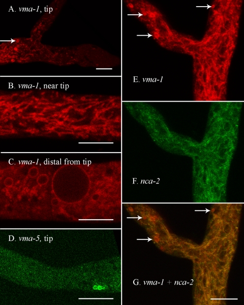
FIG. 6.
The vacuolar ATPase appears in the vacuolar network, in the membrane of larger spherical vacuoles, and in spherical organelles near the hyphal tip. The scale bar is 10 μm in all panels. (A) Tip of a hypha transformed with rfp::vma-1+. An arrow points to one of several spherical organelles that can be seen near the tip in this strain. (B) A region approximately 200 μm from the hyphal tip in a strain transformed with rfp::vma-1+. (C) A region approximately 2 mm from the hyphal tip in a strain transformed with rfp::vma-1+. (D) The tip of a hypha transformed with vma-5+::sgfp. (E to G) A region approximately 100 μm from the hyphal tip in a heterokaryon formed by coinoculating strains transformed with nca-2+::sgfp and rfp::vma-1+. Panels E and F were merged in panel G. Arrows point to red spherical organelles that can be seen near the tip.

