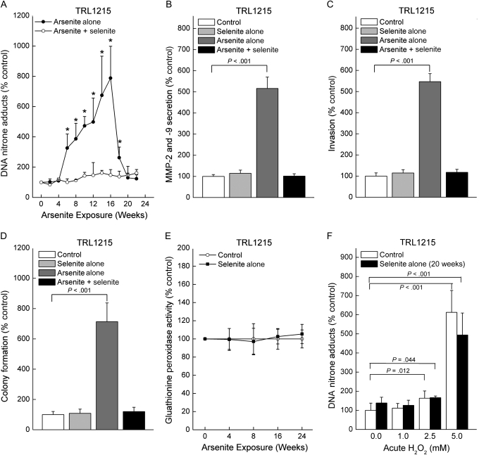
Figure 3.
Influence of exposure to sodium selenite (1.0 μM) on arsenite-induced (0.5 μM) oxidative DNA damage (ODD) and acquired in vitro cancer phenotype in TRL1215 cells. A) ODD in cells exposed to sodium arsenite in the presence or absence of sodium selenite for up to 24 weeks, as assessed by the immuno-spin trapping method. Control cells and those grown in the presence of selenite alone showed no ODD and are omitted for clarity. B) Influence of selenite on secreted matrix metalloproteinase (MMP)-2 and -9 activity induced by chronic arsenite exposure at 18 weeks, the time of malignant transformation as assessed by arsenic-induced xenograft tumor formation in a prior study (7). Assays were performed as in Figure 1, E. C) Influence of selenite on invasiveness induced by 18 weeks of chronic arsenite exposure. Assays were performed as in Figure 1, C. D) Influence of selenite on colony formation induced by 18 weeks of chronic arsenite exposure. Assays were performed as in Figure 1, D. E) Influence of selenite on glutathione peroxidase activity for more than 24 weeks of exposure. F) Influence of 20 weeks of chronic selenite treatment on hydrogen peroxide–induced ODD. Hydrogen peroxide was added to cells at indicated levels for 24 hours before ODD measurement. Untreated cells served as a control. Results in all cases are expressed as the mean with 95% confidence intervals of nine samples from three independent experiments. In panel (A), an asterisk indicates a statistically significant (P < .05) difference from control. All P values are from two-sided tests. P values were calculated by using analysis of variance followed by Dunnett multiple comparison test (B–D) or Student t test (A and F).

