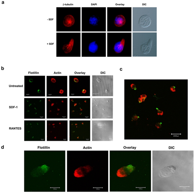
Figure 1. Pre-assembled platforms of flotillins polarize to the uropods upon chemokinesis and the spatial segregation of flotillins and actin cytoskeleton during polarization.
a) T lymphoblasts were treated with SDF-1α and allowed to migrate on fibronectin. The control cells were just plated on fibronectin without any chemoattractant. The cells were then fixed and stained with β-tubulin (red) and DAPI (blue) to stain the microtubules and the nucleus, respectively. Note that during chemokinesis, the microtubules and the MTOCs reorient themselves to the rear end of the cell. b) Untreated T lymphoblasts show uniform actin distribution but very polarized flotillin-1 staining (green). But chemoattracted (SDF-1α or RANTES) treated cells show polarized morphology accumulating actin (red) at the leading edge and flotillins (green) at the uropod. The images represent several sets of experiments and images collected. c) Magnified image of an SDF-1α treated lymphocyte displaying a clear spatial distribution of flotillins and actin cytoskeleton. d) Note that, even before a morphologic polarization took place, flotillins and actin spatially distribute themselves as early as 5 min after stimulation.

