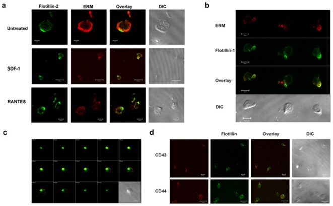
Figure 2. Flotillins co-localize with uropod markers upon chemokinesis in T lymphoblasts.
a) SDF-1α or RANTES treated lymphoblasts were stained with anti-flotillin-2 (green) and anti-ERM (red) antibodies. DIC images show morphological polarization during migration. b) ERM (red) and flotillin-1 (green) stained RANTES treated lymphoblast show that both the molecules concentrate at the uropod during migration. Note that ERM staining is also found at the leading edge due to the fact that the antibody also recognizes ezrin, which is localized at the leading edge. c) Peripheral T lymphoblasts were treated with RANTES, fixed and stained for flotillins (green) and ERM (red) proteins. Series of Z-stack images show that flotillins are more contained to the uropods. d) Chemokine stimulated cells were allowed to migrate on fibronectin and stained with anti-CD43 (upper panel, red), CD44 (lower panel, red) and anti-flotillin antibodies (green). The DIC images show the contrast image of the polarized lymphoblasts.

