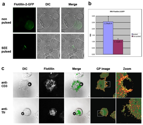
Figure 6. Flotillin polarization to the immunological synapse is concomitant with membrane condensation.
a) Jurkat cells stably expressing Flotillin-2-GFP were incubated with either unpulsed or SEE-pulsed EBV-B cells for 20 min. Confocal images were taken with identical settings. b) Quantitation of the conjugate formation in non-pulsed and SEE-pulsed conditions. c) Jurkat cells were labeled with Laurdan, conjugated with anti-CD3 antibody- or anti-TfR antibody-coated beads on ice and activated for 10 min at 37°C. After fixation, cells were immunostained for flotillin-1, adhered to poly-L-lysine-coated coverslips, mounted and imaged. The confocal image of flotillin-1 is recorded at the identical focal depth as the Laurdan images that are converted into GP images as described in Methods. GP images are pseudocolored with high GP (ordered membranes) in yellow and low GP (fluid membranes) in green.

