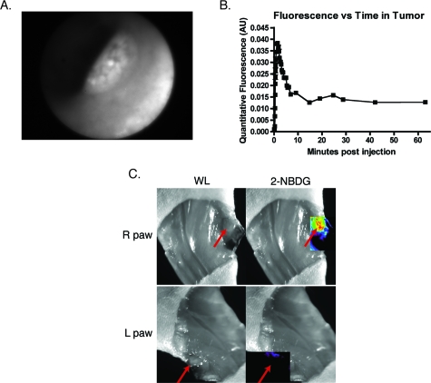
Figure 4.
Minimally invasive and intraoperative applications of 2-NBDG imaging. (a) and (b) depict data from a quantitative fluorescence colonoscopy performed on a mouse with chemical carcinogen-induced orthotopic colorectal adenomas. (a) White light (WL) image of an adenoma that was imaged using the quantitative catheter-based imaging system for one hour continuously, following the administration of 500 nmol of 2-NBDG. (b) Plot of 2-NBDG signal over time in a single adenoma shows an initial peak followed by a steady plateau. The peak likely represents 2-NBDG initially within the tumor vasculature, and the subsequent plateau reflects true accumulation and trapping of 2-NBDG within the tumor cells. (c) Epifluorescence imaging of popliteal lymph nodes in a mouse injected with JHU-31 metastatic prostate cancer cells in its right hind paw, 30 min after the administration of 500 nmol of 2-NBDG. Red arrows indicate the location of the lymph nodes. The lymph node ipsilateral to the tumor is noticeably larger in the WL image than the contralateral node; fluorescence imaging shows increased 2-NBDG uptake within the lymph node, a finding suggestive of metastatic spread.

