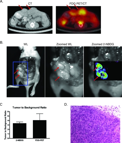
Figure 5.
Correlation of FDG-PET imaging with intraoperative 2-NBDG imaging. HT29 cells were surgically implanted into the peritoneal wall of (n=5) nude mice. The mice subsequently underwent imaging with FDG-PET∕CT, and then two days later, underwent surface reflectance imaging 30 min following the administration of 500 nmol of 2-NBDG and surgical exposure of their abdomen. (a) FDG-PET∕CT reveals two large intraperitoneal masses, shown with red arrows, that are highly FDG-avid in this representative mouse. (b) The same two lesions were well visualized with 2-NBDG imaging. Red arrows demonstrate the location of the tumors, and the blue box in the left-most image shows the approximate area of focus for the center and right-most images. (c) TBRs for FDG-PET and 2-NBDG imaging were statistically similar. (d) The malignant nature of the lesions was confirmed by histology.

