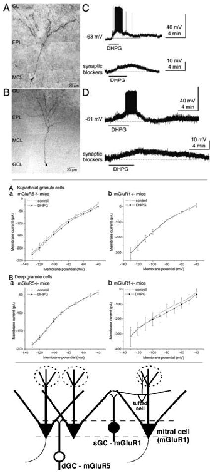
Figure 2.

Deep and superficial GCs differentially express mGluR1 and mGluR5. Upper four panels: The group I mGluR agonist DHPG (50 μM) depolarized and increased spike firing in superficial GCs (sGCs) and deep GCs (dGCs) in olfactory bulb slices from wild-type mice. Photomicrograph montage of a biocytin-filled sGC (A) and dGC (B); note soma location in the mitral cell layer (MCL) for the sGC. GCL, granule cell layer; GL, glomerular layer; EPL, external plexiform layer. (C) Bath application of DHPG depolarized and increased the discharge of an sGC (upper trace). Lower trace: DHPG applied in the presence of blockers of fast synaptic transmission (CNQX, APV, gabazine) resulted in smaller membrane depolarization in the same sGC. (D) DHPG also depolarized and increased the firing of dGCs (upper trace). Lower trace: DHPG applied in the presence of blockers of fast synaptic transmission resulted in smaller depolarization of the same dGC. Middle four panels: (A) I–V curves for sGCs in slices from mGluR5−/− (a) and mGluR1−/− mice (b), before (control, open circles) and during bath application of DHPG (50 μM, solid circles). Plots represent group data (mean ± SEM) from four sGCs (a) and five sGCs (b). DHPG evoked a current in mGluR5−/− (a) but not mGluR1−/− (b) mice. (B) Similar experiments for dGCs in slices from mGluR5−/− (a) and mGluR1−/− (b) mice. No DHPG-induced current was recorded from dGCs in slices from mGluR5−/− mice, whereas a robust current was observed in slices from mGluR1−/− mice. The inward current of dGCs in slices from mGluR1−/− mice diminished near the estimated K+ equilibrium potential (−96 mV). DHPG evoked a significant inward current (P < 0.05 compared with control) at −90 mV and more positive voltages. All experiments were performed with blockers of fast synaptic transmission and TTX in the bath. Lower illustration: MOB circuit, depicting mGluR1 expression by mitral cells and sGCs and mGluR5 expression by dGCs. Superficial dendritic extensions of deep and superficial GCs interact with different portions of M/T cell lateral dendrites. Adapted from Ref. 36.
