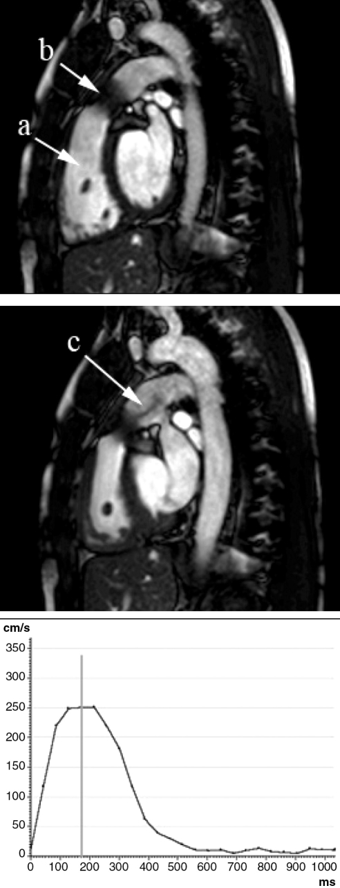
Figure 2).
Assessment of valvular stenosis. Modified sagittal end-diastolic (top) and end-systolic (middle) dynamic images illustrating right ventricle (a) and percutaneously implanted pulmonary bioprosthesis (b). Acceleration jet in the pulmonary artery (c) indicates persisting gradient measured by a velocity-encoded magnetic resonance velocity graph (bottom)

