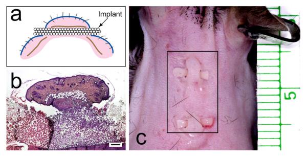
Figure 3.
Mouse implant model. (a) Cartoon of the mouse model. Blue line, epidermis; brown line, panniculus carnosus; pink, dermis. (b) low-power, H&E-stained light micrograph of a sagittal section of implant with skin above and below. Mag bar = 250 μm. (c) Photograph of implants in dorsal skin of mouse. Plate (b) from Isenhath SN, et al., J Biomed Mater Res A 83A:919, 2007, with permission from the journal.

