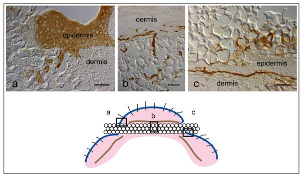
Figure 4.
Mouse implant immunomorphology. 40 μ pore diameter porous poly(HEMA) rod harvested 7 days after implant into a mouse. (a) Pan-keratin stain (Dako Corp, Carpinteria, CA), (b) Vessels [Platelet/ Endothelial cell adhesion molecule-1 (PECAM-1), Research Diagnostics, Concord, MA], (c) Laminin 332 (laminin 5) stain (courtesy of Dr. William Carter, Fred Hutchinson Cancer Research Center, Seattle, WA). The boxes in the cartoon below indicate the areas illustrated. Mag bar = 50 μm.

