
Fig. 2.
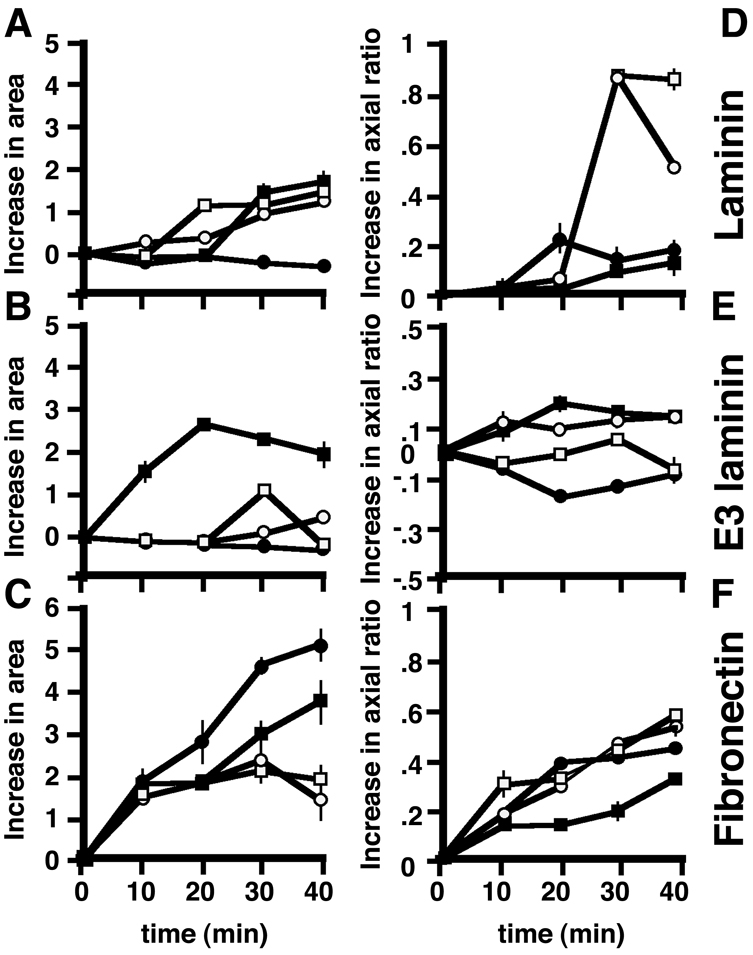
Effect of extracellular matrix and cellular dystroglycan content on cell spreading. Wild-type H2k myoblasts (white squares) or H2k myoblasts expressing control sense shRNA construct (white circles), an shRNA directed against dystroglycan (black circles) or dystroglycan-GFP (black squares) were allowed to adhere and spread for up to 40 minutes on whole laminin (A), rE3 fragment of laminin (B) or fibronectin (C). Spreading (cell area) was determined by measuring cell area using ImageJ. By 30 minutes, all cells have spread on laminin with the exception of the dystroglycan-knockdown cell line. On the rE3 fragment of laminin, only cells overexpressing dystroglycan are able to spread. Interestingly, on fibronectin, cells depleted for dystroglycan spread best of all. (D-F) Change in axial ratio with time for the same groups of cells as measured in A-C. Cells were assumed to be round at time 0 (whether they were or not), and all measurements of axial ratio calculated on that basis. Hence some cells, especially those on E3 show a negative value, because the cells became even rounder over time. All cells polarise well on fibronectin (F) and poorly on rE3 laminin (E). On laminin, cells with altered levels of dystroglycan do not polarise well, whereas controls on laminin are the most polarised of all (D).