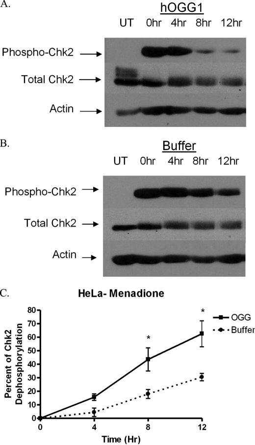
FIGURE 8.
MTS-hOGG1-Tat mediates inactivation of Chk2. HeLa cells were pretreated with the hOGG1 fusion protein and then exposed to menadione (200 μm). The results show that hOGG1 (A) can decrease phosphorylation of Chk2 (Thr-68) more rapidly than buffer-treated controls (B), thereby demonstrating an effect resulting from enhanced mitochondrial BER. UT, samples untreated by menadione. Times shown are the number of hours following menadione treatment that the cells were harvested. C, quantitation of the density of the bands from the resulting films showing a statistical change in the phosphorylation state in the hOGG1-treated samples compared with buffer-treated controls (n = 3).

