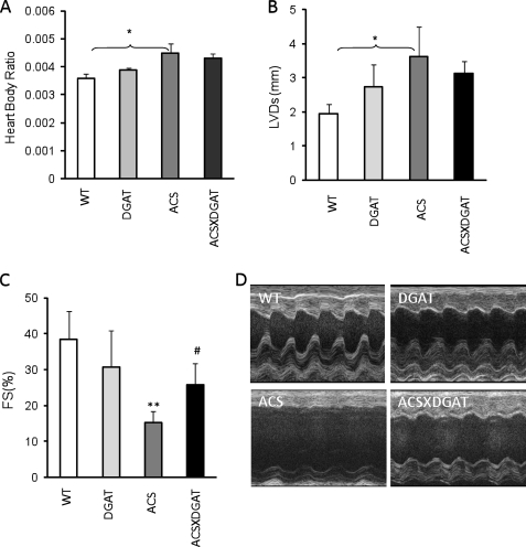
FIGURE 4.
Cardiac function in MHC-DGAT1 and MHC-DGAT1/MHC-ACS mice. A, heart to body weight ratio in WT, MHC-DGAT1, MHC-ACS, and MHC-DGAT1/MHC-ACS transgenic mice (n = 5–7). Echocardiography showed left ventricular systolic dimension (B) and fractional shortening (C). Photographs are shown of echocardiograms (D) (n = 5–7). **, p < 0.01 MHC-ACS versus WT or MCH-DGAT1; #, p < 0.05 MHC-ACS versus MHC-ACS/MHC-DGAT1.

