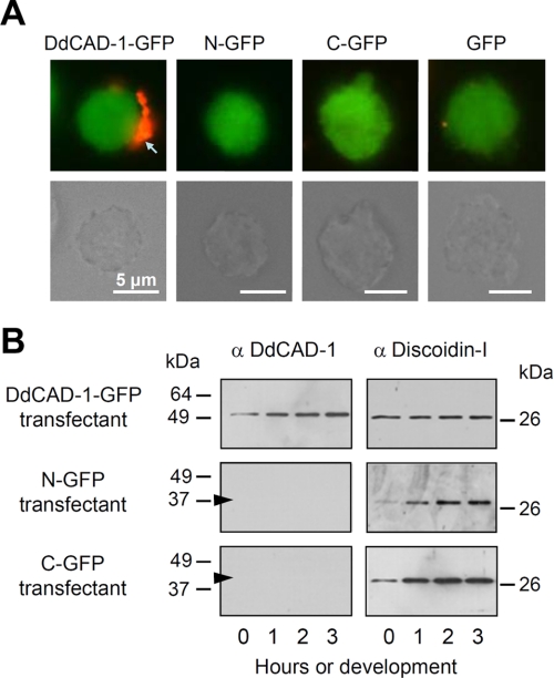
FIGURE 4.
Differential cell surface expression and secretion of the DdCAD-1 fusion proteins in transfectants. A, fluorescence micrographs showing antibody-induced cap formation of DdCAD-1-GFP on live cells. cadA-null cells expressing the different GFP fusion proteins were incubated with anti-GFP antibodies for 30 min. After washing, cells were incubated at room temperature for another 30 min with a secondary antibody to induce “cap” formation (red). The corresponding light micrographs are shown in the lower panels. Bars, 5 μm. B, DdCAD-1 secretion during development. Transfectants were developed in 17 mm phosphate buffer at 2 × 10⁷ cells/ml, and the conditioned media were collected at 1-h intervals for Western blot analysis using rabbit antibodies against DdCAD-1 or discoidin-I. The arrowheads indicate the position of N-GFP (38 kDa) and C-GFP (41 kDa), respectively.

