
Figure 5.
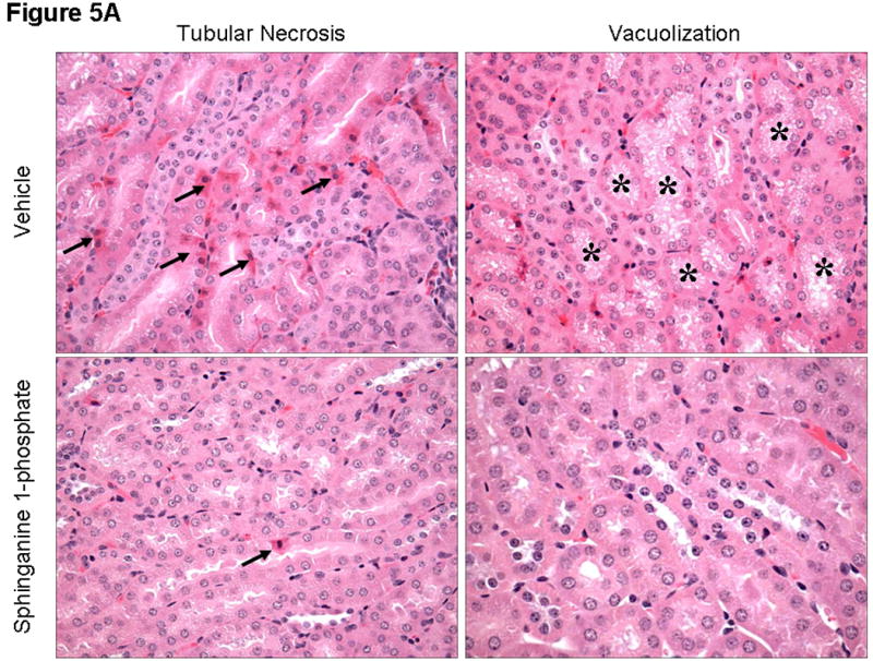
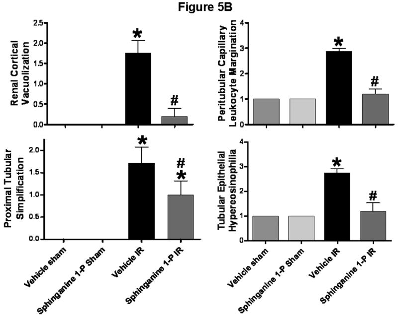
A). Representative photomicrographs of 6 experiments (hematoxylin and eosin staining, magnification 400×) demonstrating hypereosinophillia (arrows) and vacuolization (*) in kidneys from C57BL/6 mice 24 hrs after being subjected to liver ischemia reperfusion after vehicle- or sphinganine 1-phosphate treatment (0.1 mg/kg i.v. immediately prior to reperfusion and 0.2 mg/kg s.c. 2 hrs after reperfusion). B). Summary of renal injury scores (scale 0-3) for renal cortical vacuolization, peritubular leukocyte margination, proximal tubule simplification and renal tubular hypereosinophilia for kidneys from mice subjected to liver IR after vehicle- or sphinganine 1-phosphate treatment. Data are presented as means ± SEM. *P<0.05 vs. Vehicle sham group. #P<0.05 vs. Vehicle IR group.