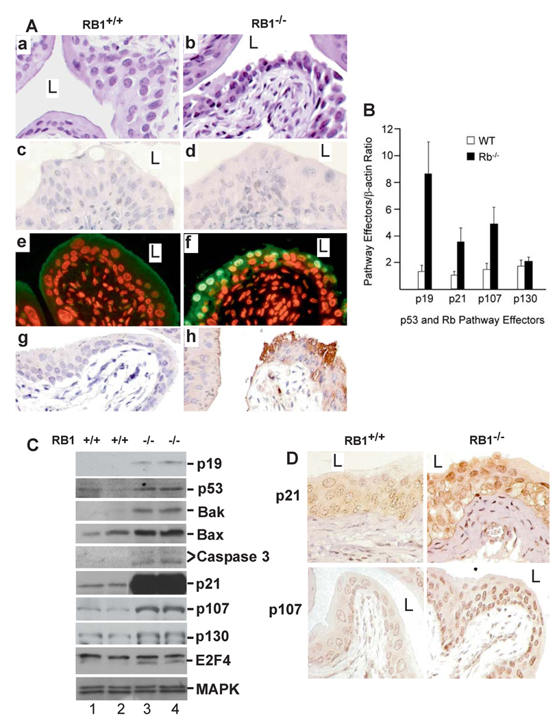
Figure 2. Urothelial response to pRb deficiency.
(A) H&E-stained urothelia of (a) an RB1+/+ mouse (15-month old) showing normal morphology and (b) an age-matched, RB1−/− mouse showing condensed nuclei and widened intercellular space. (c & d) anti-Ki67 staining showing lack of urothelial proliferation in the RB1−/− mouse. (e & f) TUNEL assay revealing apoptosis in the RB1−/− mouse (f) but not in the RB1+/+ control (e). (g & h) anti-caspase 3 antibody strongly labeling urothelial cells in RB1−/− mouse (h) but not in the RB1+/+ mouse (g). Magnification: 200 ×. (B–D) Real-time RT-PCR (B), Western blotting (C) and immunohistochemistry (D) showing strong induction of p53 pathway components p19, p53, Bak, Bax, activated caspase 3 fragments, p21 and pRb family member p107 and its binding partner E2F4, but not p130, in RB1−/− mice. n=8 in (B).

