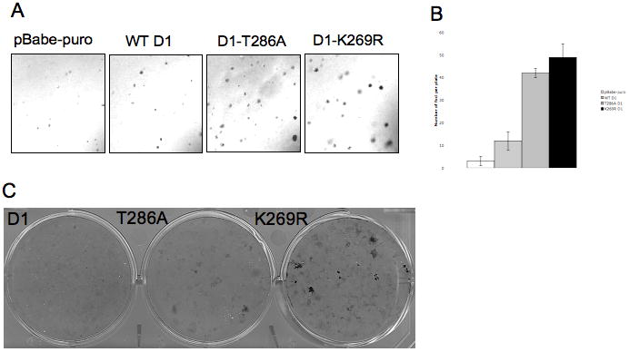
Figure 5. K269R cyclin D1 transforms NIH-3T3 cells.

A. Anchorage –independent growth of NIH-3T3 cells stably expressing WT cyclin D1, D1-T286A, D1-K269R or pBabe-puro vector was analyzed by growth in soft agar. Colonies were visualized by microscopy. B. Quantification of triplicate samples shown in A. C. Foci formation ability of NIH-3T3 cells stably expressing WT cyclin D1, D1-T286A and D1-K269R was determined by Giemsa stain of foci grown for 21 days.
